 |
47 (9) (1995), pp. 13-17. JOM is a publication of The Minerals, Metals & Materials Society |
|---|
 |
47 (9) (1995), pp. 13-17. JOM is a publication of The Minerals, Metals & Materials Society |
|---|
The single-user nature of traditional SEMs also affects the manner in which they are utilized in industrial or research settings, where access is typically restricted to a small number of individuals. This may be due to a number of factors. Examples include the lack of trained personnel or lack of time available to train large numbers of people, the cost associated with training since typically only one or two people can be trained at a time, the physical separation of the SEM from the potential users, the difficulty of mastering the instrument itself, and/or the desire to prevent damage to the instrument by allowing unqualified users to operate the machine. Single-user or restricted-user facilities are the norm in almost all cases. Such arrangements often prevent individual engineers and/or researchers from personally examining samples or checking the results of samples examined by technicians.
The problems associated with both scenarios can be overcome by adopting a new method for SEM instruction and operation, one that allows multiple users access to the instrument in a pseudo-parallel, rather than sequential, manner. This necessitates developing an SEM that gives multiple users complete and independent control of the SEM and allows them to access and analyze SEM images and data in a parallel manner.
A first step in developing such a system has been taken at Iowa State University, where a conventional SEM has been connected to a network of computer-based remote workstations to provide students with direct, interactive access to the SEM. Control of the microscope is achieved by means of a local-area network (LAN), which also allows the students access to image analysis software and x-ray energy-dispersive spectroscopy (EDS) capabilities. The results thus far have exceeded expectations, and it is evident that possible applications of such a network exist in both industrial and research areas.
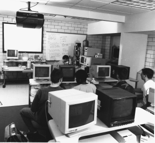
Figure 1. Photograph of a multi-user SEM classroom. At this time, four remote stations are in operation with plans to expand to six.
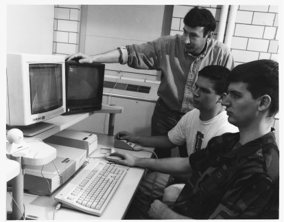
Figure 2. Photograph of a typical remote teaching station, consisting of a computer, video monitor, thermal printer, and joystick control. This station also contains a small camera for video conferencing.
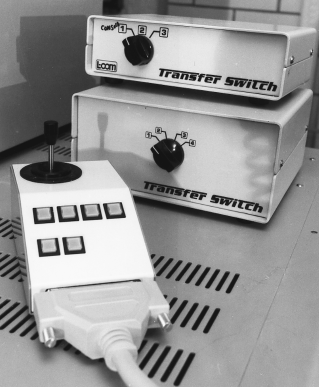
Figure 3. Close-up of a typical joystick control built at Iowa State University to mimic the microscope joystick and the switcherbox system.
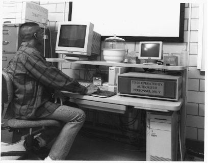
Figure 4. The main computer control station for the laboratory consists of an Apple Quadra equipped with 40 Mb of RAM, a 500 Mb hard drive, and boards that provided EDS control, Ethernet connection, frame-grabbing capabilities, a modem, and four additional serial ports. The station also holds hardware used in operating the EDS system.
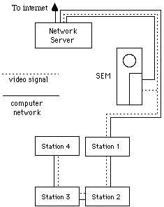
Figure 5. Schematic showing the layout of the laboratory and the local area network used.
The microscope is controlled by a program called Scope,2 which was written at Iowa State University. Scope allows the control of various operating parameters on the microscope and has been designed to be adaptable to more advanced models or machines from different manufacturers. The main control screen from Scope is shown in Figure 6. The JEOL 6100 only allows limited access to the control parameters of the microscope, requiring selection of accelerating voltage, beam saturation, and all initial alignment steps to be done from the microscope console. However, through Scope students can change focus, magnification, and probe current, and have access to other options such as auto brightness and contrast, the text editor, the scan coils, and the shutter used to take photographs. Scope only resides on the network server and is only available to the student using Timbuktu in the control mode.
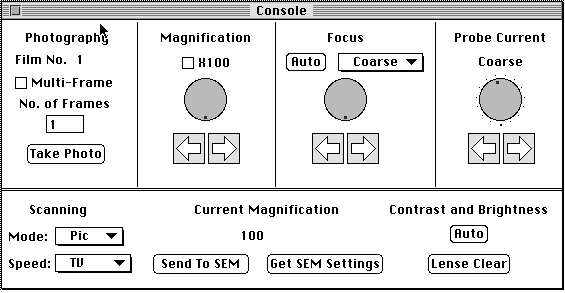
Figure 6. The console view from the program Scope developed at Iowa State University to control the microscope.
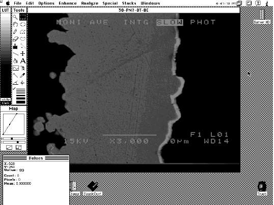
Figure 7. A typical screen from the program Image.
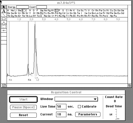
Figure 8. A typical screen from the program Microplus.
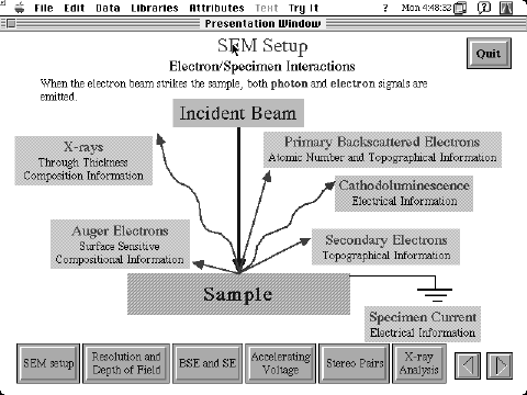
Figure 9. A typical screen from the SEM tutorial program written at Iowa State University.
The combination of these software packages enables all students to be involved at all times when not actively operating the microscope. When control of the microscope is desired, it is achieved simply and quickly with the click of a mouse. Thus, instruction is enhanced and the productivity of the lab period is increased.
Iowa State University possesses an extensive computer network system, providing Ethernet access to the Internet's World Wide Web and connectivity to almost any place on campus. This allows access to the SEM laboratory on a 24 hour basis from essentially any networked computer on or off campus. This flexibility is utilized in a number of ways. Students are able to access the network directly from home and do not have to return to the laboratory to retrieve data or images stored on the remote stations. The instructor is able to monitor and control the server from both office and home computers by using Timbuktu. Student operation of the server computer can be viewed and override commands issued if desired. The sample can be viewed over the Ethernet connection by acquiring the microscope image using the frame grabber board and Image shareware. Thus, the instructor can monitor student operation without being present in the classroom.
The disadvantage of using a network software control rather than a hardware control in order to operate the microscope is related to the response speed of the system. Although this is not a serious shortcoming, remote operation using Timbuktu is slower than direct operation of the microscope from each station via a dedicated line. A recent upgrade to Timbuktu Pro® has greatly increased the speed of the network, improving the operation of the network considerably; however, if an even faster response is desired, speed may be further increased by having Scope reside on each remote station. A direct connection of each station to the RS-232 port of microscope via a switcher box arrangement similar to that used for the joystick control enables the instructor to switch control between stations. This situation might be appropriate for a small classroom setting.
The great advantage of using software, however, is that it makes operation and control of the microscope possible from any location that possesses the necessary software. It is this flexibility that raises interesting possibilities for the extension of the multi-user concept to industry and research environments.
Since the software-controlled concept allows the SEM to be operated from any suitably equipped computer on the Internet, links have been established with the local high school to provide students in physics, biology, and chemistry with the opportunity to see a sample in near-live time from the high school classroom. Small digital cameras in the classroom and the SEM laboratory allow video conferencing between the high school students and the SEM operator. The Timbuktu network software allows high school students to observe the SEM image and EDS spectra collection on their computer screen. These initial results prove that the multi-user SEM concept can be extended from the laboratory to essentially any location.
As part of this project, a page is being developed for the World Wide Web that will allow interested persons to download various SEM images of insects, plants, and materials. Another planned feature will carry a video image of the SEM, allowing viewers to watch students and researchers as they conduct their investigations.
A multi-user SEM may have numerous applications in areas besides education. For example, we expect that the system could operate in a research facility. While a technician would still be responsible for inserting the sample into the microscope, individual scientists could access the SEM from their respective offices. The scientists could perform the actual investigation or simply monitor the results seen by the technician. The results could be viewed immediately by a whole team of researchers either in their offices or in a conference room setting. Control of the microscope could be passed between investigators quickly and simply, allowing different samples, or different regions of a single sample, to be viewed in turn. In any case, the results of the analysis would be known instantly, and pictures and data could quickly be incorporated into a document or transferred electronically to other researchers.
A similar arrangement could be considered in an industrial setting. Production and quality control personnel could be linked directly to the company SEM facility, allowing them to view images and examine samples without leaving their work areas. Potential problems identified by the SEM operator could immediately be communicated to engineers responsible for production. The engineer could examine the suspect part personally, pass the information and image on to other involved personnel, and take immediate steps to rectify any problems. Such an arrangement will speed information transfer and increase productivity.
There would be no need for extensive training in these scenarios since the software controls could be limited to simple stage movement and control of magnification and focus. Instruments are now on the market that allow computer control of these parameters; thus, there would be very little training involved for the user before they could take advantage of the most basic capabilities of the microscope. Any potential user could be easily taught the keyboard controls necessary to operate the microscope, obtain the necessary data, and pass it along to relevant personnel.
Another advantage is that time is not lost in transit to the location of the SEM since a computer screen and keyboard can be placed almost anywhere. In fact, the SEM need not even be at the plant site, but could be anywhere in the world that has access to the Internet.
There is no barrier to the widespread adoption and usage of multi-user SEM microscopy that cannot be solved by careful planning and a willingness to adopt modern computer-based methods of operation.
ABOUT THE AUTHORS
L.S. Chumbley earned his Ph.D. in metallurgy at the University of Illinois in 1986. He is currently an associate professor at Iowa State University. Dr. Chumbley is also a member of TMS.
M. Meyer earned his Ph.D. in ceramic engineering at Iowa State University. He is currently a post-doctoral fellow at Ames Laboratory, Iowa State University. Dr. Meyer is also a member of TMS.
K. Fredrickson earned his B.S. in computer engineering at Iowa State University in 1994. He is currently a software engineer at MTS in Eden Prairie, Minnesota.
F.C. Laabs earned his M.S. in physics at Mankato State University in 1965. He is currently an associate metallurgist at Iowa State University.
For more information, contact Scott Chumbley, Ames Laboratory, Iowa State University, 214 Wilhelm, Ames, Iowa 50011; (515) 294-7903; fax (515) 294-4291; e-mail: chumbley@iastate.edu.
Direct questions about this or any other JOM page to jom@tms.org.
| Search | TMS Document Center | Subscriptions | Other Hypertext Articles | JOM | TMS OnLine |
|---|