An Article from the January 2003 JOM-e: A Web-Only Supplement to JOM
|
TMS
ONLINE | TMS
PUBLICATIONS | SITE
MAP An Article from the January 2003 JOM-e: A Web-Only Supplement to JOM |
|
|
|
|
Jeongguk
Kim and Peter K. Liaw are with the Department of Materials Science and
Engineering at the
University of Tennessee. H. Wang is with
Oak Ridge National Laboratory. J.Y. Huang and R.C. Kuo are with the
Institute of Nuclear Energy Research in Taiwan. J.G. Huang is with
the
Taiwan Power Company.
|
Exploring traditional, innovative, and revolutionary issues in the minerals,
metals, and materials fields.
|
| OUR LATEST ISSUE |
|
|
OTHER ARTICLES IN THE SERIES |
|---|
|
|
|
In 1829, the first fatigue test was performed in Germany on steel
chains, which were subjected to 100,000 tension cycles at a frequency of ten
cycles per minute. Since then, a great amount of valuable research effort has
been devoted to studying the fatigue behavior of materials due to the irreplaceable
significance of this subject in the engineering world.1–8
In recent decades, fracture mechanics has become the dominant concept used in
describing and understanding fatigue behavior.
Although there is still interest in such concepts as fracture toughness and
endurance limit, data scattering and time-consuming, costly experiments can
cause difficulties during fatigue testing of industrial components and structures.
Thus, increasing efforts have been focused on nondestructive evaluation (NDE)
techniques for their critical importance in fatigue-life assessments, structural-integrity
evaluations, failure prevention, and material savings. Several NDE methods have
been applied to monitor mechanical damage, including ultrasonics, acoustic emission,
eddy current, x-ray, and computed tomography,9–12
Relatively little work has been done to characterize fatigue behavior using
thermographic infrared techniques.13–17
In this paper, a study has been conducted on the background and theory of thermography.
Furthermore, an advanced high-speed and high-sensitivity infrared (IR) imaging
system was used to monitor the temperature evolution of reactor pressure vessel
steels subjected to 10 Hz, 20 Hz, and 1,000 Hz fatigue tests. Both thermodynamics
and heat-conduction theories are applied to explain and model the observed temperature
evolutions during fatigue and to predict the inelastic behavior.
What Is Thermography?
Thermography is a nondestructive evaluation (NDE) technique that monitors the
target temperature change. Originally, the technique was used primarily in the
military service to observe enemy movement at night and in hospitals to monitor
the temperature change of organs and tissues. Later on, as the relationship
between mechanical properties and temperature was better clarified and the technique
became more advanced, thermography developed into an important nondestructive
detection technique in the engineering world. The relationships between the
materials temperature evolution and mechanical behaviors can be explained in
light of three effects:
|
The History of Thermography in Materials Science
The relationship between temperature and material deformation was recognized
in 1853 by Kelvin18 and
then developed by Biot,19
Rocca, and Bever20 in
the 1950s. In the 1960s, Dillon21
and Kratochvil22 developed
the thermoplastic theory that directly relates the temperature with the material
internal stress-strain state, which, in turn, controls the mechanical and fatigue
behavior. In 1956, Belgen23
developed IR radiometric techniques for detecting temperature changes. These
techniques calculated temperature change with stress by measuring the IR radiation
emitted from the surface of solid materials. However, the limited sensitivity
of the available instrumentation restrained the full development of thermography
as a NDE technique until the 1980s, when rapid developments in electro-optical
and signal processing techniques led to a more advanced IR imaging system, an
IR camera.24–26
With a temperature resolution up to 10–3°C
and a spatial resolution up to several micrometers, this new equipment makes
possible the practical quantitative stress-strain analysis by temperature. Recent
research has shown the potential of thermography in monitoring mechanical damage.27–40
However, detailed investigations and comprehensive analyses are needed to develop
a more practical thermography method in characterizing the fatigue process.
Material
The material used in fatigue tests is reactor pressure vessel (RPV) steel (SA533B1I2),
which is composed of, in weight percent, 0.203C, 0.23Si, 1.34Mn, < 0.02P,
0.015S, 0.50Ni, 0.53Mo, 0.15Al, 0.005N, 0.01Cu, and balance, Fe. The steel plate
was first solution-treated at 899°C for 1 h, then water-quenched to 40°C,
and finally tempered at 670°C for 1 h. A tempered martensite was the final
microstructure. The yielding strength of the RPV steel was 587 MPa, ultimate
tensile strength was 716 MPa, and total elongation was 29% with a strain rate
of 4 × 10⁻³/s and a gage length
of 1.27 cm used in the tension test. A yielding-point phenomenon was observed
in the test.40
Fatigue Testing
Fatigue test samples are cylindrical bars with a gage length of 1.27 cm and
a diameter of 0.508 cm at the gage section. The test samples are machined from
the steel plate with the length direction parallel to the rolling direction
and then polished in a sequence of 240, 400, 600, and 800 grit papers, followed
by 9.5 mm, 1 mm, and 0.06
mm Al2O3
grit powders.
The 1,000 Hz fatigue tests were performed using an advanced high-frequency electrohydraulic
Material Test System (MTS) machine (Model 1,000 Hz 810) with an R ratio of 0.2,
where R = smin/smax,
and smin and smax
are the applied minimum and maximum stresses, respectively. The servovalves
of the machine were activated by voice coils, which provide the necessary frequency
of 1,000 Hz. The machine has a maximum loading capability of ±25 KN.
The test samples were fastened to the machine by specially designed mechanical
grips. To avoid the testing noise at 1,000 Hz, the machine was situated in a
well-designed soundproof room equipped with a heat pump that offered the cooling
capability to prevent the overheating of servovalves.
For 10 Hz and 20 Hz fatigue experiments in air, the specimens were loaded on
a MTS machine (Model 810) at R = 0.2. A load control mode was used, and different
maximum stress levels ranging from 500 MPa to 650 MPa were applied. During fatigue
testing, an extensometer, which was connected with the data-acquisition system
of the MTS machine, was fixed on the gage-length section of the specimen. The
strain values of the gage length, measured by the extensometer, could then be
recorded directly into the MTS system for further analyses.
In addition, the tensile properties of the RPV steel were determined using a
plate sample that was 10 mm in width, 35 mm in length, and 5 mm in thickness
of the gage-length section. The tensile test was performed at a strain rate
of 5 × 10⁻⁴/s.
Thermography
Thermography detection was conducted using a state-of-the-art Raytheon Galileo
thermographic IR imaging system with a 320 × 256 pixel focal-plane array
InSb detector that is sensitive to a radiation wavelength of 3 mm
to 5 mm. The temperature sensitivity is 0.015°C
at 23°C, while the spatial resolution can be as small as 5.4 mm.
The system’s maximum-speed data acquisition capability is 120 Hz at a
full frame of 320 × 256 pixels and 50,000 Hz at 16 × 16 pixels.
During fatigue testing, a thin sub-micrometer graphite coating was applied on
the specimen gage-length section to decrease the surface-heat reflection.
A thermocouple was attached to the sample to calibrate the IR camera at the
beginning of each test. During calibration, the specimen was heated to a high
temperature with a heat gun, then air cooled. The temperature of the specimen
was recorded from the thermocouple at different times, and the intensity of
the IR camera was calibrated to the corresponding temperature. A fully automated
software system was employed to acquire the data of temperature distributions
of the test samples during fatigue experiments. The IR camera was used at low
speeds of 0.1 Hz and 0.2 Hz, and a high speed of 120 Hz.
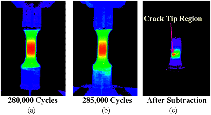
Figure 1 exhibits the IR images of RPV steels at a maximum stress of 600 MPa, R-ratio of 0.2, and test frequency of 1,000 Hz, which were taken at an IR camera speed of 1 Hz. Figure 1a presents a temperature profile of the fatigued sample at 280,000 cycles, while Figure 1b shows a temperature profile at 285,000 cycles. In both Figure 1a and 1b, the highest temperature region is located in the gage-length section of the specimen, as represented in red color. Subtracting the temperature distribution at 280,000 cycles from that at 285,000 cycles indicates the occurrence of cracking (Figure 1c). The red color in the subtraction image (Figure 1c) identifies the presence of a single hot spot. This is believed to be located at a crack tip, where the heat generation is the greatest. In that location, a significant amount of plastic deformation occurs, which becomes the heat source. Similar results can be found at 20 Hz. Thus, thermography can be used to identify the presence of crack initiation and propagation during fatigue testing.
|
|
 |
|
Figure 1. IR images of RPV specimen high-cycle, fatigue-tested at 1,000 Hz, smax = 600 MPa, and R = 0.2. |
|
|
| |
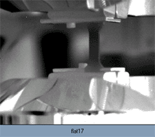 |
| Animation 1. A movie depicting the Lüders band evolution process of RPV specimen during 10 Hz fatigue testing. |
| |
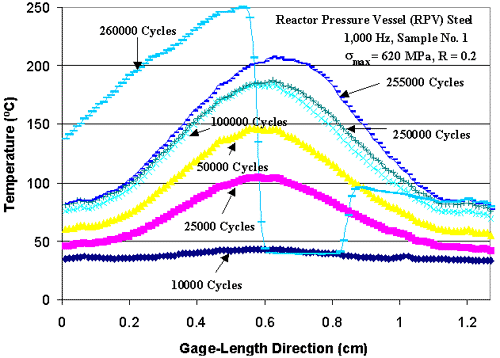 |
| Figure 2. A temperature line profile of a RPV specimen during 1,000 Hz fatigue testing. |
Figure 2 exhibits the average
temperature distribution along the gage-length (1.27 cm) direction of the specimen
corresponding to different cycles at a maximum stress of 620 MPa, R-ratio of
0.2, and test frequency of 1,000 Hz, taken at an IR camera speed of 1 Hz. Positions
0 and 1.27 on the x axis represent the two ends of specimen gage-length section.
The temperature was generally found to be the highest at the midpoint of the
sample. The temperature rises rapidly at first from 10,000 cycles to 100,000
cycles, and becomes stable after 100,000 cycles, then goes up sharply and quickly
to failure after 250,000 cycles. Near the center of the specimen, the temperature
can rise from about 40°C to 250°C depending on the cycles. The temperature
distribution curve at 260,000 cycles shows the temperature variation immediately
after the specimen breaks, which rises abruptly in the last 5,000 cycles, relative
to that at 255,000 cycles. At 260,000 cycles, the specimen fractures and separates,
which results in a temperature drop in the specimen gage-length section between
0.6 cm and 0.8 cm (i.e., the specimen separates between 0.6 cm and 0.8 cm in
the gage-length section).
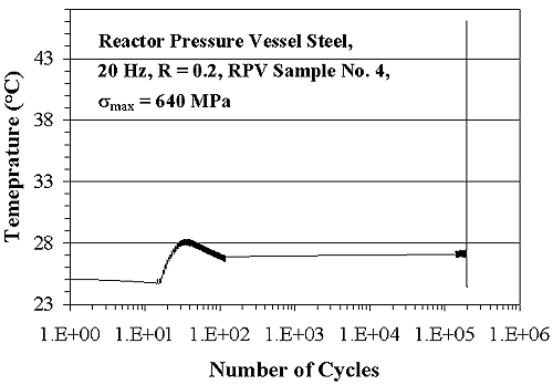
Figure 3 shows the temperature evolution,
at the mid-point of the specimen gage-length section, plotted on a log scale
of fatigue cycles for the 20 Hz fatigue test with a R ratio of 0.2 and maximum
stress level of 640 MPa. The specimen temperature at the midpoint of the gage-length
section initially increases from 23.7°C to 28.5°C with fatigue cycling,
followed by a temperature decrease (i.e., a temperature hump) in the first 100
cycles. After that, the temperature approaches a steady state of about 27°C
and then increases abruptly to 49°C until the specimen fails. A detailed
analysis of the temperature profile has been provided in previous work.35,36,38,40
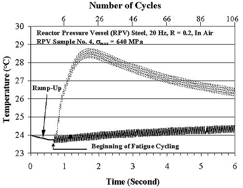
In Figure 4, the dashed line represents
the amplified hump in Figure 3. In Figure
4, at the very beginning stage, a slight temperature decrease within the
first 0.7 s was due to the thermoelastic effect, as discussed later. Then, the
temperature rose rapidly from the first fatigue cycle at approximately 0.7 seconds
and a temperature of 23.7°C, and reached a maximum of 28.5°C in about
2 s. After that, the temperature decreased gradually to a relatively constant
value. However, if the test was stopped after the temperature became stable,
and then, restarted, no temperature hump was observed. The corresponding results
are plotted as a solid line in Figure 4.
Note that in both tests shown by the dashed and solid lines, temperature oscillations
within the range of approximately less than 0.6°C were observed within each
fatigue cycle.
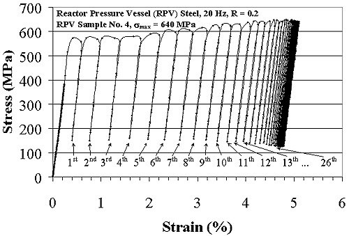
Since the mean temperature variation is closely related to the plastic deformation,21,22
a reasonable explanation of the presence of the temperature hump can be obtained
from the stress-strain curve in Figure 5.
This is a typical stress-strain curve for the tension-tension fatigue test.
Corresponding to the temperature rise from approximately 0.7 s to 2 s in Figure
4, the stress-strain curve in Figure
5 moves from the first cycle to the 26th cycle, and the plastic strain increases
from 0 to nearly the saturated value of about 4.7%. In this period, a great
amount of heat is generated from the large plastic deformation and the temperature
of the sample increases quickly. Moreover, the yielding-point phenomenon of
RPV steels is observed in the uniaxial tensile test, which contributes to large
plastic strains (Figure 5) and, in turn,
more heat is generated.
However, after the first 26 cycles, relatively little plastic strain occurs
due to the strain-hardening effect in Figure
5, and the temperature decreases when the heat inside the sample is conducted
to the environment, finally reaching a relatively constant value due to the
heat equilibrium between the heat generation of the specimen subjected to cyclic
loading and the environment. Note that, in Figure
5, the maximum stress level is lower than 640 MPa for the first several
cycles. This trend results from the fact that the fatigue machine needs some
time to reach a stabilized stress level at the beginning of fatigue testing.
|
|
|
|
||
 |
 |
 |
||
|
Figure 3. The specimen temperature evolution of reactor pressure vessel steel during 20 Hz fatigue testing, taken at an IR camera speed of 120 Hz. |
Figure 4. The temperature-versus-time evolutions of reactor pressure vessel steel tested at 20 Hz, taken at an IR camera speed of 120 Hz. |
Figure 5. The stress-versus-strain result of reactor pressure vessel steel tested at 20 Hz, smax = 640 MPa. |
||
|
|
|
|
If the fatigue test is terminated and restarted, little heat will be generated from the plastic deformation since the plastic strain has already saturated. Thus, there will be no rapid temperature rise in the first 100 cycles, as indicated by the solid lines in Figure 4. The stress-strain curve of the restarted test is exhibited in Figure 6, which presents much less plastic deformation as compared to Figure 5, resulting in a much smaller temperature rise shown in the solid line, relative to the dashed line in Figure 4. Thus, there is a good correspondence between the temperature evolutions and the stress-strain characteristics during fatigue.
| |
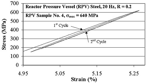 |
| Figure 6. The stress-versus-strain profiles of reactor pressure vessel steel tested at 20 Hz, smax = 640 MPa. |
| |
This project is supported by the Taiwan Power Company. The authors are also very grateful for the financial support of the National Science Foundation (DMI-9724476, EEC-9527527, and DGE-998-7548) with D.R. Durham, M.F. Poats, W. Jennings, and L. Goldberg as contract monitors, respectively, and the Tennessee Advanced Materials Laboratory with E.W. Plummer as the director. A portion of the work was sponsored by the U.S. Department of Energy Secretary for Energy Efficiency and Renewable Energy, Office of Transportation Technologies, as part of the High Temperature Materials Laboratory User Program under contract DE-AC05-96OR22464, managed by the UT-Battelle.
References
1.
S. Suresh, Fatigue of Materials, 2nd Ed. (Cambridge, U.K.: Cambridge
University Press, 1998).
2. W.A. Logsdon, P.K.
Liaw, and J.A. Begley, ASTM
STP 969 (1988), pp. 830–867.
3. P.K. Liaw, W.A.
Logsdon, and J.A. Begley, Metallurgical
Transactions, 20A (1989), pp. 2069–2085.
4. P.K. Liaw and W.A.
Logsdon, J.
Engineering Materials and Technology, 107 (1985), pp. 26–33.
5. P.K. Liaw et al.,
Engineering
Fracture Mechanics, 57 (1997), pp. 85–104.
6. W.A. Logsdon and
P.K. Liaw, Engineering
Fracture Mechanics, 22 (1985), pp. 509–526.
7. J.Y. Huang et al.,
“Fatigue Behavior of Reactor Pressure Vessel Steels,” Julia Weertman
Symposium, ed. Y.W. Chung et al. (Warrendale, PA: TMS,
1999), pp. 373–384.
8. J.Y. Huang et al.,
“Fatigue Behavior of SA533-B1 Steels,” ASTM
STP 1406 (Philadelphia, PA: ASTM,
2001), pp. 105–121.
9. L. Jiang et al.,
Nondestructive
Evaluation (NDE) and Materials Properties IV, ed. P.K. Liaw (Warrendale,
PA: TMS, 1999), pp. 43–60.
10. M.E. Fine, Z.M.
Connor, and J.D. Achenbach, Nondestructive
Evaluation (NDE) and Materials Properties IV, ed. P.K. Liaw (Warrendale,
PA: TMS, 1999), pp. 1–9.
11. G. Birnbaum
and G. Free, “Eddy-current Characterization of Materials and Structures:
A Symposium Sponsored by ASTM Committee E-7 on Nondestructive Testing (American
Society for Testing and Materials, Gaithersburg, MD, 5–7 September
1979).
12. A.A. Moss and
H.I. Goldberg, Computed Tomography, Ultrasound and X-ray: An Integrated Approach
(New York; Masson Publication, 1979).
13. M.P. Luong,
Mechanics
of Materials, 28 (1-4) (1998), pp. 155–163.
14. K.S. Hermanson
and B.I. Sandor, Experimental Techniques, 22 (3) (1998), pp. 19–21.
15. X. Tung, D.
Wang, and H. Xu, Acta Metallurgica Sinica (China), 28 (4) (1992), pp.
A163–169.
16. D. Zhang and
B.I. Sandor, ASTM STP 1122
(1991), pp. 341–353.
17. P.K. Liaw et
al., editors, Nondestructive
Evaluation (NDE) and Materials Properties IV (Warrendale, PA, TMS, 1999).
18. I. Todhunter
and K. Pearson, A History of the Elasticity and Strength of Materials, Vol.
2 (Cambridge, U.K.: Cambridge
Press, 1893).
19. M.A. Biot, J.
Appl. Phys., 27 (3) (1956), pp. 240–253.
20. R. Rocca and
M.B. Bever, Trans. Am. Inst. Mech. Eng., 188 (February 1950), pp. 327–333.
21. O.W. Dillon,
J. Mechanics and Physics in Solids, 11 (1963), pp. 21–23.
22. J. Kratochvil
and O.W. Dillon, J. Applied
Physics, 40 (8) (1969), pp. 3207–3218.
23. M.E. Belgen,
J. Applied Physics,
27 (3) (1956), pp. 240–253.
24. D.H. Allen and
W.E. Haisler, Mechanics and Structures, 13 (1981), pp. 129–135.
25. E.G. Henneke,
K.L. Reifsnider, and W.W. Strinchcomb, J.
Metals, 31 (1979), pp. 11–15.
26. R.H. Blanc and
E. Giacometti, “Infrared Radiometry Study of the Thermomechanical Behavior
of Materials and Structures” (Paper presented at the First International
Conference of Stress Analysis by Thermoelastic Technics, Sira Ltd, London, November
1984).
27. Y. Frum, Int.
J. Pres. Ves. & Piping, 61 (1995), pp. 367–381.
28. P. Stanley and
W.K. Chan, J. Strain Analysis, 20 (3) (1985), pp. 129–137.
29. G. White and
G. Torrington, Material Evaluation (1995), pp. 1332–1335.
30. U. Hansen, J.
Composite Materials, 33 (7) (1999), pp. 614–639.
31. S. Offermann
et al., Experimental Mechanics, 37 (1997), pp. 409–413.
32. J. Roth, J.R.
Bodis, and C. Bishop, NASA Technical Memorandum 106950 (1995).
33. B. Nayroles
et al., Int. J. Eng. Sci., 19 (1981), pp. 929–947.
34. D.T. Lohr, N.F.
Enke, and B.I. Sandor, Dynamic Failure: Proceedings of the 1987 SEM Fall
Conference (Bethel, CT, Soc.
for Experimental Mechanics, 1987), pp. 169–174.
35. P.K. Liaw et
al., Scripta
Materialia, 42 (2000), pp. 389–395.
36. H. Wang et al.,
Metall. and Mater.
Transactions A, 31 (2000), pp. 1307–1310.
37. M.P. Luong,
Mechanics
of Materials, 28 (1998), pp. 155–163.
38. L. Jiang et
al., Transactions of Nonferrous Metals Society of China, 12 (2002), pp.
734–747.
39. H. Wang et al.,
Metallurgical and Materials
Transactions A, 33 (2002), pp. 1287–1292.
40. B. Yang et al.,
Materials Science and Engineering, A314 (2001) pp. 131–139.
For more information, contact P.K. Liaw, University of Tennessee, Department
of Materials Science and Engineering, 427-B Dougherty Engineering Building,
Knoxville, TN 37996-2200; e-mail pliaw@utk.edu.
Direct questions about this or any other JOM page to jom@tms.org.
| Search | TMS Document Center | Subscriptions | Other Hypertext Articles | JOM | TMS OnLine |
|---|