 Presenting a Web-Enhanced Presenting a Web-Enhanced Feature Article from JOM |
 LATEST ISSUE |
|||
TMS QUICK LINKS: |
• TECHNICAL QUESTIONS • NEWS ROOM • ABOUT TMS • SITE MAP • CONTACT US |
JOM QUICK LINKS: |
• COVER GALLERY • CLASSIFIED ADS • SUBJECT INDEXES • AUTHORS KIT • ADVERTISE |
|
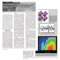
| Overview: Neutron Scattering | Vol. 58, No.3, pp. 47-51 |
Structure Determination and Phase
Analysis by the Use of Neutron Diffraction
Simon J.L. Billinge
| ||||||||||||
| ||||||||||||
| ||||||||||||
|
| ||||||||||||
|
Questions? Contact jom@tms.org. © 2006 The Minerals, Metals & Materials Society |


|
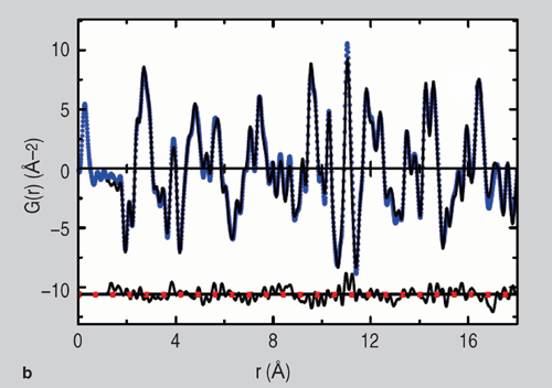
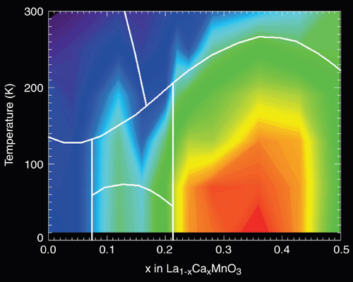
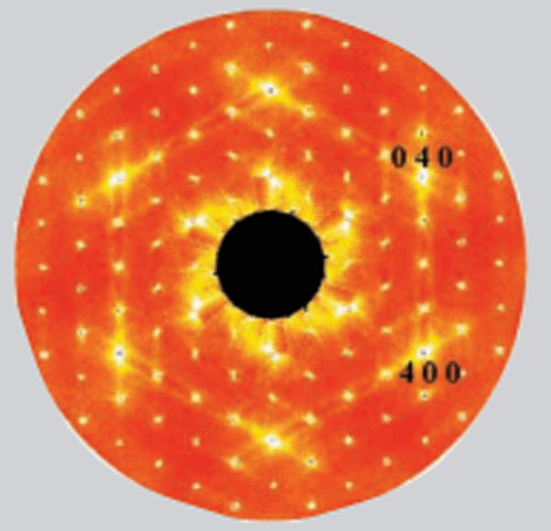
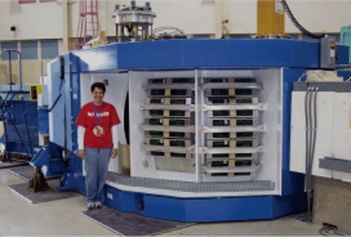
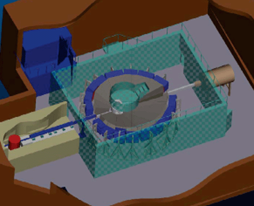
INTRODUCTION When William Lawrence and William Henry Bragg looked around their laboratories in 1912 as they did their Nobel-prize-winning research, using x-rays to determine atomic structure, the materials they saw would not have been so remarkably different from those in Michael Faradays lab 50 years earlier, or indeed Isaac Newtons 150 years before that: wood, leather, glass, brass, and steel. The pace of change in materials science has dramatically accelerated in the past 100 years, resulting in a wealth of diverse and useful novel materials. The timing is not completely coincidental, as knowledge of atomic-scale structure underpins our understanding of material properties and our ability to design new materials. One hundred years later, x-ray diffraction remains one of the most important tools in materials science, so ubiquitous that it hardly needs introduction to this audience.1 Atomic structures are solved from single-crystal data and structural parameters, accurate to hundredths and even thousandths of an angstrom. They are routinely determined by profile refinement of single crystals and powder data.2-4 As sources get more powerful and computers get faster, the complexity of structures that can be solved ever increases such that protein structures with thousands of unique atomic positions can be determined. Structural databases now contain hundreds of thousands of distinct structures.5 Parametric studies as a function of composition, temperature, and pressure allow detailed determinations of phase diagrams, and data of high precision and resolution allow defects and disorder to be studied in crystalline materials. Peak searching algorithms linked to databases seek candidates for constituents of powdered phase mixtures.6 For disordered materials, which present a particular challenge in the detailed understanding of their structure, diffraction is an irreplaceable component in their study.7 Finally, diffraction is playing a key role in nanotechnology, where it is desired to control structure, and therefore properties, of materials on the nanoscale.8,9 What is the role for neutron diffraction in this larger picture? X-rays would seem to have two main advantages: accessibility and intensity. Virtually every materials lab has access to some kind of x-ray diffractometer locally at the institution, giving excellent access to x-rays. On the other hand, in experiments where high intensity is of great importance, those with tiny samples, for example, can be taken to x-ray synchrotron sources with unparalleled flux and brilliance. Despite these factors favoring x-rays, neutron diffraction has proven to be an indispensable and highly significant tool since its development 60 years ago. The neutron has unique properties that yield distinct information unavailable otherwise. Chief among these properties is the relative uniformity of the neutron scattering power of atoms across the periodic table, allowing the accurate structure determination of materials composed of widely differing atomic-number species.10 Among the most highly cited papers of all time in materials science are those discussing structures of high-temperature superconductors and colossal magnetoresistant manganites that were made possible by neutron powder diffraction.11 The activity of these materials depends sensitively on the oxygen positions that are virtually invisible in an x-ray experiment due to the proximity of strongly scattering lanthanides. The relatively strong scattering of neutrons off magnetic moments also ensures that the majority of magnetic structures are solved using neutron diffraction.12,13 The high penetrability of neutrons in matter, due to their lack of charge, is helpful for studies on samples in special environments, in-situ studies, and very large samples, such as structural components, as described in the article in this issue by Xun-Li Wang on engineering diffraction. Materials with significant disorder, from glasses to disordered crystals via nanoparticles, are studied using total scattering methods where both Bragg and diffuse scattering intensities are analyzed on an equal footing.8,9 This approach was developed to study glasses and liquids in the early days of diffraction.14 However, coupled with powerful sources and fast computers, these approaches, such as the atomic pair distribution function (PDF) method, are finding much wider application. The PDF method involves a Fourier transform of the data from reciprocal-space to real-space. An example of a PDF, the fit, the data, and the structure that was fit, is shown in Figure 1. To obtain a useful resolution in real-space, data must be collected over a wide range of momentum transfer, Q, and another property of the neutron is useful here. The neutron is scattered by the atomic nucleus, which is a point on the length-scale of the neutron wavelength (1015 m compared to 1010 m). As a result, there is no Q-dependent form-factor that kills the coherent scattering intensity at high scattering angles as with x-rays, making these high-resolution PDF measurements straightforward with neutrons. The scattering power also depends on the actual isotope as well as the chemical nature of the scatterer. This property is utilized to gain chemical insight in hard structural problems. By taking the difference between measurements from two samples that are chemically identical, but where the isotope of a particular species has been enriched in one of the samples, the structure of a chemical sublattice of the total structure can be determined. This is particularly important in disordered materials and is widely used there;16,17 however, it can also provide crucial input in complex crystallographic problems.18,19 This is similar to the use of anomalous scattering in x-ray diffraction experiments. The experiments are considerably more straightforward, though more pricey. One final very elegant twist to this tale is to utilize the incoherent scattering properties of the neutron. Bragg peaks and all the structural information require coherent scattering. As mentioned previously, in neutrons the scattering power depends on the particular isotope. Additionally, it depends on the relative spinstates of the nucleus and the neutron interacting with it. These properties change from neutron to neutron and site to site in a crystal in a random way, leading to an incoherent scattering component in neutron scattering that is not present with x-rays. Normally this incoherent scattering is an annoying background in diffraction experiments. However, at particular, fortuitous compositions it is possible to suppress individual Bragg peaks completely, allowing delicate higher-order effects in the material to be studied.20 Null-scattering alloys, such as a vanadium-niobium alloy, can also be made with no coherent scattering whatsoever, despite being crystalline; these are useful as sample containers in neutron diffraction experiments. NEUTRON DIFFRACTION EXPERIMENTS Whilst neutron diffraction experiments cannot be conducted in the laboratory (they need a high flux source of neutrons), they are easy to carry out for anyone familiar with x-ray diffraction. Once you have the data, similar, and often the same, refinement programs that are used for x-ray diffraction refinements are also used to refine the neutron data. Indeed, after entering the experimental parameters, the refinements proceed in an identical manner. It is possible, and can be desirable, to co-refine x-ray and neutron datasets within the same program, such as with the program General Structure Analysis System.21 It should be noted, too, that Rietveld refinement, the ubiquitous powder-diffraction profile-fitting model refinement method, was originally invented by Hugo Rietveld22 for reactor neutron data that have very beautifully compliant Gaussian peak shapes. This is much easier to handle than laboratory or synchrotron x-ray data. For newcomers, there are two main obstacles to carrying out neutron experiments. The first challenge is to obtain samples of sufficient size for effective neutron experiments (although with modern sources this obstacle is rapidly being removed since data rates allow x-ray-sized samples to be studied.) The second obstacle is access to a neutron diffractometer. This is often the greatest psychological obstacle for those not familiar with the process but it is straightforward and, apart from travel expenses, the beam time is free for non-proprietary research. Typical beam-time allocations at reactor and spallation national user facilities are for 17 days at a time and these are allocated by a proposal system on scientific merit. Information about the facilities and the proposal application system can be found at the facility web sites. A list and links to neutron facilities and user groups can be found under the links tab of the Neutron Scattering Society of America (www.neutronscattering.org). Data from a single sample at a single temperature take from a few minutes to a few hours to collect, depending on the scattering properties of the sample and the properties of the diffractometer chosen. For room-temperature data from a few samples, a number of facilities are now offering rapid access mail-in services. One can simply ship samples to the facility, which will run them and send the data back. This is an excellent way to get your feet wet and to see if neutron data will be of use in your research. The proposal preparation process takes some getting used to (you may scratch your head at a few of the questions at first) and by far the gentlest introduction to this process for someone who has never tried it before is to find someone to collaborate with, though this is not a requirement. If you dont have a pet neutron scatterer nearby then a good approach is to e-mail one of the instrument scientists at the facilities. I would be delighted to put you in contact with someone who may be able to help. Types of Science Powder measurements are carried out on a wide range of materials. As mentioned, neutrons have a huge advantage for finding lighter atoms in the presence of heavy atoms in the structure, and a significant amount of work has been carried out on oxides, sulfides, and so on. Locating hydrogen in structures and determining magnetic structures is also important in neutron powder work. Despite having relatively high throughput for neutron instruments, powder diffractometers are in high demand and generally have among the highest over subscription rates, which is a testament to the power and usefulness of this technique. The experiments are straightforward and rapid, allowing parametric studies (Figure 2), and the analysis software is rather mature, opening the technique to a large community. Less mature, but no less important, are studies of disorder in crystalline materials and structure solution of nanostructured materials. Neutron diffraction is set to have a large impact here, too, for the same reasons as in crystalline materials: the complementarity of neutron and x-ray data. Nanocrystalline materials yield broad and poorly defined scattering peaksthere is some discussion in the field as to whether we should refer to them as Bragg peaksmaking traditional Rietveld refinements of limited value. However, novel total scattering approaches, such as atomic pair distribution function analysis8,9 where the data are Fourier transformed to real-space and the real-space profile is refined, are showing great promise (Figure 1). Ab-initio structure solution of clusters and molecules in real-space has also recently been demonstrated. The demonstration was carried out on neutron data from C60 buckey balls.26 As our thrust toward controlling matter on the nanoscale intensifies, these techniques look set to become of increasing importance in materials science. Disorder in crystals can also be studied from the diffuse scattering in single crystals. Spallation neutron single-crystal diffractometers such as SXD at the ISIS Pulsed Neutron & Muon Source (ISIS) in Chilton, United Kingdom, collect full segments of reciprocal space as a matter of course, making them particularly attractive for this kind of measurement. An example of diffuse scattering from disordered benzil crystals is shown in Figure 3. Types of Diffraction Instruments In the early days of spallation sources, the odd diffraction line shapes led to lower-quality refinements than from reactors. This is no longer the case with improvements in the modeling programs and the choice between reactors and spallation sources now comes down to other factors. For single-crystal measurements, there are only a handful of diffractometers to choose from around the world. On the other hand, powder diffractometers tend to be somewhat optimized for particular measurements and there are a number of choices to make. Magnetic structure determinations and atomic structure refinements on materials with large unit cells need a diffractometer with good flux and high resolution at low scattering angles. Total scattering and PDF measurements need a wide range of momentum transfer. In these measurements, good flux at high-Q are a necessity, so they are most practical at spallation sources. Parametric studies and studies on small samples need high flux and are better carried out on moderate resolution, high-flux instruments. Most neutron instruments will accommodate various special environments including (moderate) high-pressure equipment, furnaces, and low-temperature refrigerators and cryostats, which are provided by the facilities, but the availability of a special environment may be a factor in choosing the instrument to use. In general, it is useful to contact one of the instrument scientists and start asking questions; you are likely to get good advice and enthusiastic encouragement, even if you end up on a different instrument because of the nature of your experiment. THE FUTURE The future for neutrons looks bright. Europe has made strong investments in neutron scattering with a new target station at the excellent ISIS spallation source (www.isis.rl.ac.uk) that will almost double their capacity, upgrades to the best research reactor in the world at Institute Laue Langevin (www.ill.fr), and continual instrument and detector development there. In addition, new sources are either under construction or recently operating in Switzerland and Germany. Japan is building a next-generation spallation (http://j-parc.jp/index-e.html) source to rival the Spallation Neutron Source (SNS) in the United States, and Australia is about to start up a new research reactor (www.ansto.gov.au/opal). There is widespread appreciation around the world that neutron scattering will be an essential tool of the 21st century. A more complete list of neutron scattering research facilities around the world is available in the links section of www.neutronscattering.org. In the United States, there is a similar optimism. The SNS (www.sns.gov), which will produce its first neutrons in 2006, is an ambitious next step in neutron sources that is set to increase the power, and therefore neutron flux, over existing sources with state-of-the-art instrumentation. In the diffraction suite, this will host a powder diffractometer, POWGEN3, of unprecedented power (Figure 5); NOMAD, an optimized high-flux disordered materials diffractometer; and SNAP, a powder diffractometer optimized for high-pressure studies. There will be two single-crystal diffractomers, TOPAZ and MANDI, for small and large unit cell structures, respectively. This is the initial suite and all are funded and under construction. More beam ports are available for future innovations in instrument design. This complements existing facilities including the excellent reactor user facilities at the National Institute of Standards and Technology (www.ncnr.nist.gov) and Oak Ridge (http://neutrons.ornl.gov) and spallation sources at Los Alamos (http://lansce.lanl.gov) and Argonne national laboratories (www.pns.anl.gov). New, more powerful, data-analysis software is an integral part of carrying out science at a facility such as this. The early (Nobel-prize-winning) experiments of Cliff Shull in the 1940s and 1950s had a single detector and could be analyzed by hand. POWGEN3 will produce roughly 1 gigabyte of data per hour from its large banks of highly pixilated detectors. Software is being developed for this purpose (e.g., see the DANSE project at http://wiki.cacr.caltech.edu/danse/index.php/Main_Page). The conjunction of modern powerful neutron instruments with software promises to result in not only more science and better science, but qualitatively new science. It is up to us to utilize these tools for qualitatively new materials science. Join the fun. ACKNOWLEDGEMENTS I would like to thank Jason Hodges for the engineers rendition of POWGEN3 and Thomas Proffen for the photo of NPDF. I would also like to thank all my hard-working students and post-docs over the years, and last, but by no means least, the patient and tireless instrument scientists and technicians at the neutron facilities. Work in the Billinge group is supported through National Science Foundation grants CHE-0211029 and DMR-0304391 and Department of Energy grant DE-FG02-97ER45651. REFERENCES 1. A.D. Krawitz, Introduction to Diffraction in Materials Science and Engineering (London: Wiley, 2001).2. C. Hammond, The Basics of Crystallography and Diffraction (Oxford, U.K.: Oxford University Press, 2001). 3. W.I.F. David et al., Structure Determination from Powder Diffraction Data (Oxford, U.K.: Oxford University Press, 2002). 4. V. Pecharsky and Peter Zavalij, Fundamentals of Powder Diffraction and Structural Characterization of Materials (Amsterdam: Springer-Verlag, 2005). 5. See www.ccp14.ac.uk/database.htm for a list of useful crystallographic-related databases. 6. The International Center for Diffraction Data (www.icdd.com/) maintains the database of diffraction patterns used by most (if not all!) peak-search programs. 7. A.C. Wright, Glass Physics and Chemistry, 24 (1998), pp. 148179. 8. S.J.L. Billinge and M.G. Kanatzidis, Chem. Commun.(2004), pp. 749760. 9. T. Egami and S.J.L. Billinge, Underneath the Bragg Peaks: Structural Analysis of Complex Materials (New York: Pergamon, 2003). 10. www.ncnr.nist.gov/resources/n-lengths. 11. For example, R.J. Cava et al., Physica C, 165 (1990), pp. 419433; J.D. Jorgensen et al., Phys. Rev. B, 41 (1990), pp. 18631877; H.Y. Hwang et al., Phys. Rev. Lett., 75 (1995), pp. 914917; P.G. Radaelli et al., Phys. Rev. B, 55 (1997), pp. 30153023. 12. Y.A. Izyumov, V.E. Naish, and R.P. Ozerov, Neutron Diffraction of Magnetic Materials (Amsterdam: Springer-Verlag, 1991). 13. J. Rodriguez-Carvajal, Physica B, 192 (1993), pp. 5569. 14. B.E. Warren, X-Ray Diffraction (New York: Dover, 1990). 15. Th. Proffen et al., Phys. Rev. B, 60 (1999), p. 9973. 16. I.T. Penfold and P.S. Salmon, Phys. Rev. Lett., 67 (1991), pp. 97100. 17. I. Petri, P.S. Salmon, and H.E. Fischer, Phys. Rev. Lett., 84 (2000), pp. 24132416. 18. A.M. Balagurov et al., Physica C, 228 (1994), pp. 299308. 19. P.F. Henry, M.T. Weller, and C.C. Wilson, J. Appl. Crystallogr., 34 (2001), pp. 4246. 20. J.A. Rodriguez et al., Abstract MP36 (Paper presented at the American Conference on Neutron Scattering, College Park, MD, 610 June 2004), www.ncnr.nist.gov/acns/program/abstract/mp36.html. 21. A.C. Larson and R.B. Von Dreele, General Structure Analysis System (GSAS), Report No. LAUR-86-748 (Los Alamos, NM: Los Alamos National Laboratory, 1987). 22. H.M. Rietveld, J. Appl. Crystallogr., 2 (1969), p. 65. 23. C.C. Wilson, Single Crystal Neutron Diffraction from Molecular Materials (Singapore: World Scientific, 2000). 24. D.A.A. et al., Physica B, 241 (1997), pp. 11221130. 25. K. Kurihara et al., Proc. Natl. Acad. Sci., 101 (2004), pp. 1121511220. 26. P. Juhas et al., Ab Initio Solid State Nano-structure Determination, Nature (to be published in 2006). 27. T.R. Welberry et al., J. Appl. Cryst., 36 (2003), pp. 14401447. Simon J.L. Billinge is with the Department of Physics and Astronomy at Michigan State University in East Lansing, Michigan. For more information, contact Simon J.L. Billinge, Michigan State University, Department of Physics and Astronomy, East Lansing, Michigan, 48824; email billinge@pa.msu.edu. |